
Scan
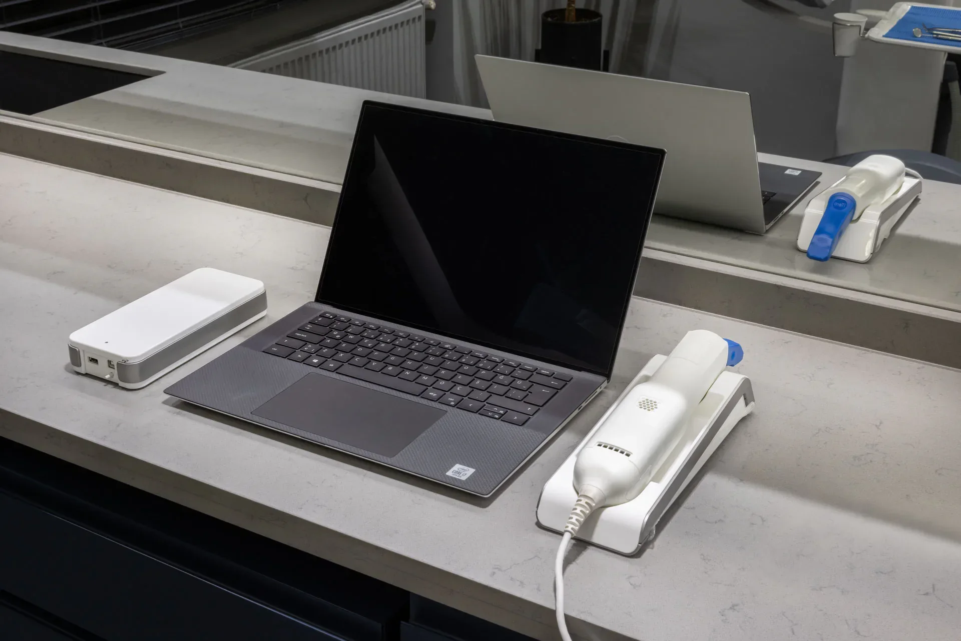
The intraoral scanner captures the teeth and gums with light and builds a 3D model on screen. No impression trays or putty are used, which many people with a strong gag reflex find more comfortable.
Intraoral scanning, 3D modelling, digital smile design and CAD/CAM: how digital tools support planning and treatment, and where their limits are.
Digital dentistry means using computer-aided tools for records, planning and the production of some restorations. These tools are not a treatment in themselves; they help make impressions, shared plans and designs more predictable.
Systematic reviews show that intraoral scanning is faster than conventional impressions, that most patients prefer it, and that accuracy is comparable for limited work such as short bridges. For some situations, such as extensive prostheses, conventional impressions may still be recommended. Which method is used therefore depends on the treatment.
We cover this in more detail in our article on intraoral scanners and digital impressions.
Each stage, from scan to bonding, on its own card.
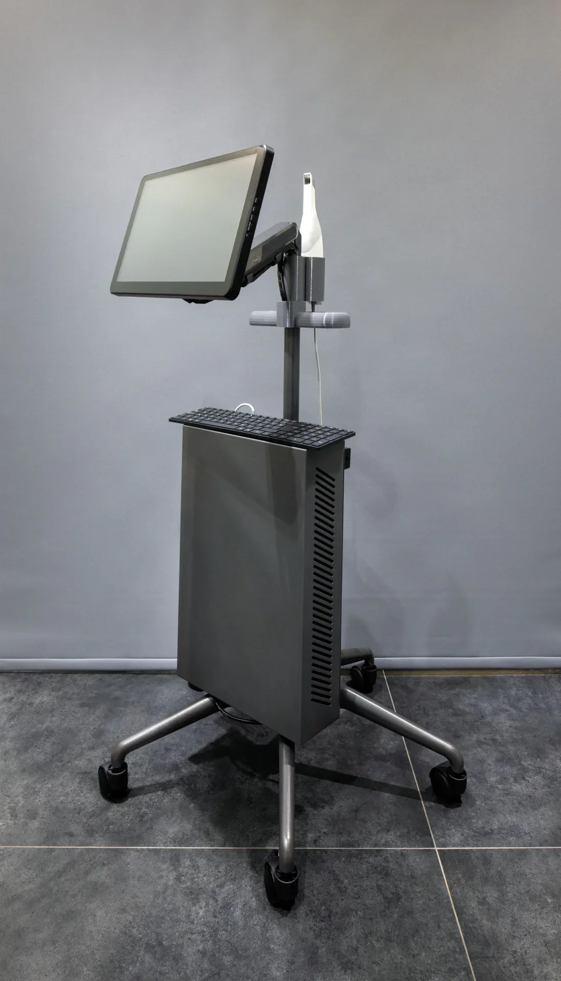
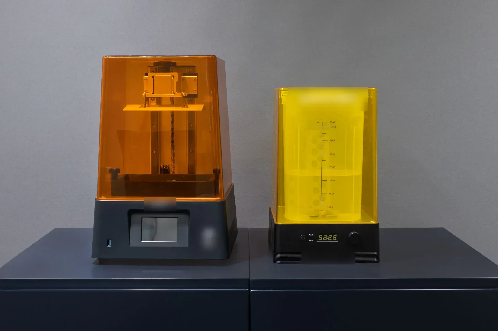
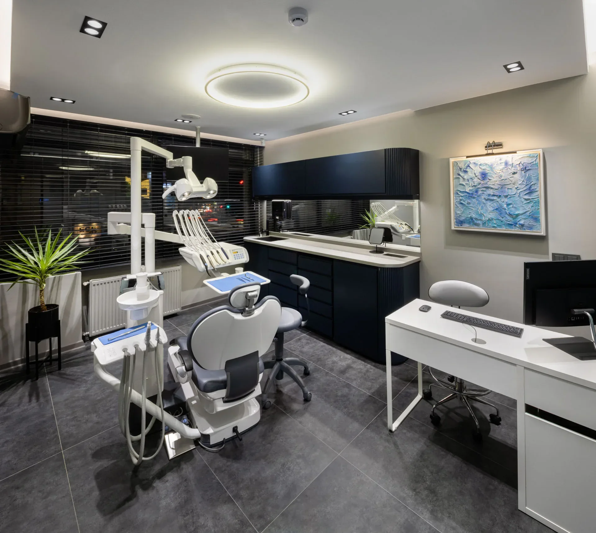
Scanning records the tooth surface optically and involves no X-rays. The resulting 3D model is the starting point for many treatments, including crowns, inlays and onlays, veneers, implant crowns, night guards and smile design.
The model is also a way of explaining: seeing the teeth and the planned change together on screen makes decisions easier, and digital records allow changes to be compared at review appointments.
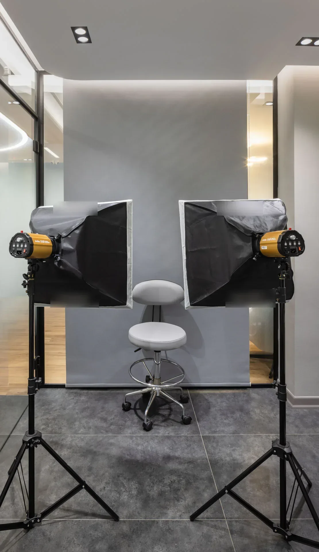
Smile planning uses facial and smile photographs taken from standard angles under controlled light. They help assess how the midline, smile line and tooth proportions relate to the face.
The digital draft built from photographs and scan data can be tried in the mouth in a temporary material when suitable. The process is described on the smile design page.
Digital tools do not help equally in every case. Conventional impressions may be preferred for some work such as extensive prostheses, and some restorations need laboratory support. A digital design is not a guarantee of the final result; materials, tooth structure and conditions in the mouth all play a part.
X-rays and 3D imaging (CBCT) are used only when needed and at the lowest suitable dose.
No. Intraoral scanners record the surface of the teeth optically using light; they do not use X-rays. X-rays are taken separately only when the inside of the teeth or the bone needs to be assessed.
Access to your personal data is one of your rights under Turkish data protection law. Ask the practice how to obtain a copy of your records; they can be useful if you continue care with another dentist.
We talk about your concerns and goals and examine your teeth and gums. X-rays are taken if needed, and when planning is involved, photographs and an intraoral scan are recorded. Options, stages and maintenance are then discussed together.
Which digital tools are used depends on your treatment and is decided after the examination.